Latest Technology in Frankfort
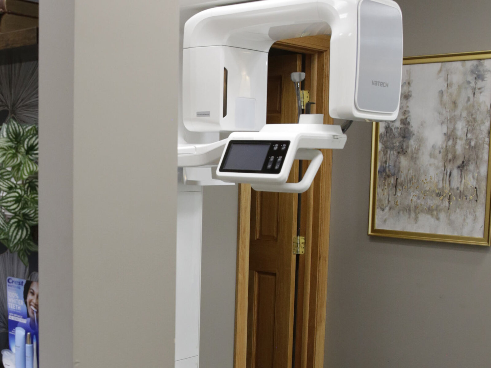
3D Cone Beam Imaging
A complete 3D view of your anatomy.
In a single, low-radiation scan, our in-office cone beam CT (CBCT) scanner builds a detailed 3D picture of your teeth, jaw, and the structures that support them. That depth of detail gives our doctors the precision they rely on for implant planning, root canal evaluation, airway assessment, and complex restorative cases.
Best of all, the imaging happens right here, so there is no separate trip elsewhere for advanced scans. We can map out your treatment and talk it through with you during the very same visit.
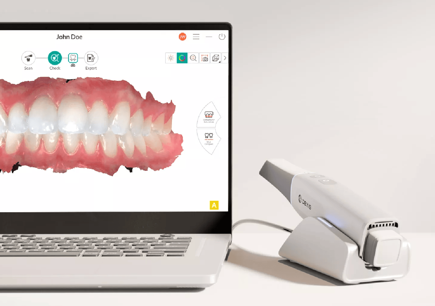
Digital Impressions: No Putty Required
Fast, precise, and completely comfortable.
Say goodbye to traditional putty impressions: the DEXIS IMPREVO is an advanced digital intraoral scanner that builds a precise 3D model of your teeth and gums instead. Small, lightweight, and wand-style, it is easy to maneuver, and most patients tell us the scan feels far more comfortable than the old impression method.
From there, the scan data flows straight into our restorative workflow, so crowns, veneers, and other restorations are designed directly from it. That removes the delays and the fit issues that tend to follow physical impressions sent to an outside lab.
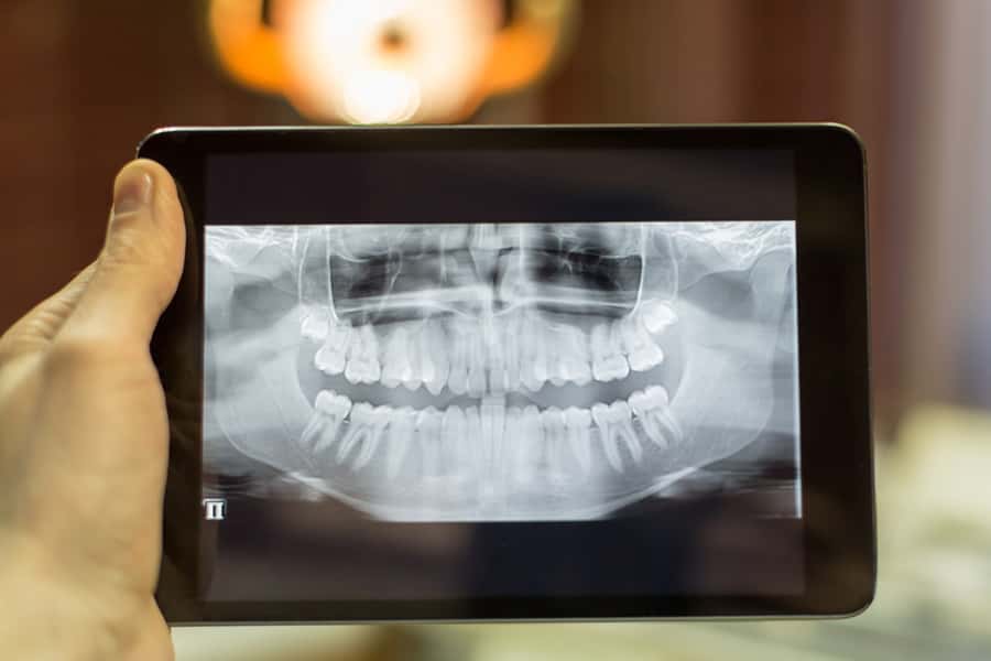
Digital Radiography
Digital x-rays are the newest technology dentistry uses to take and archive dental x-rays, and they do it while significantly reducing the amount of radiation compared to traditional dental x-rays. The technique captures a digital picture of your teeth along with their supporting bone structures and stores the images on a computer in our dental office. Within moments, you and your doctor can view your x-rays together and enlarge the image to aid in the identification of dental problems and to gauge your dental health.
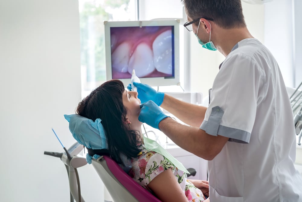
Intra-Oral Camera
About the size of a pen, the intra-oral camera is a small video camera that photographs the teeth and supporting tissue, a wonderful addition to dentistry today. It creates digital images that can be stored on a computer, and we share those images with our patients so they can join us in the "co-diagnosis" of problems with their dental health. Your doctor can show you how others view your smile and point out which dental fillings are broken or discolored.